
Testicular Ultrasound


Testicular Ultrasound
Essential information
A testicular ultrasound assesses the testicles, epididymis, spermatic cord, and surrounding tissues. It is commonly requested to investigate pain, swelling, a palpable lump, changes in testicular size, or concerns found during examination.
This scan is the primary imaging method used to detect testicular abnormalities and is a key investigation for early detection of testicular cancer.
Testicular cancer is the most common cancer in men aged 15 to 50, although it can occur at any age. The number of cases has doubled over the past 20 years, with around 1500 new cases diagnosed each year in the UK. Early detection and treatment significantly improve outcomes by reducing the risk of spread.
Ultrasound provides clear, real-time images that help identify cysts, solid masses, infections, inflammation, or other structural changes. You should consult your doctor to ensure any necessary follow-up or onward referral is arranged.
Why should I choose MedicalUltrasound.co.uk for my testicular ultrasound?
- Experienced Sonographer Led Scrotal Imaging
Your testicular ultrasound is performed by an experienced Sonographer with specialist expertise in scrotal and male pelvic imaging, ensuring accurate and sensitive assessment of the testes and surrounding structures. - Focused Testicular and Scrotal Assessment
The scan is specifically designed to assess the testes, epididymis, and scrotal contents to investigate lumps, pain, swelling, trauma, or fertility-related concerns, and to help identify conditions such as cysts, inflammation, varicoceles, or other abnormalities. - High Quality Real Time Ultrasound Imaging
We use modern ultrasound technology, including Doppler imaging where appropriate, to produce clear, real-time images that support detailed evaluation of testicular structure and blood flow. - Safe, Non-Invasive and Radiation-Free Investigation
Testicular ultrasound does not involve radiation and is non-invasive, making it suitable for urgent assessment, reassurance, and repeat imaging when required. - Clear Clinical Reporting for Onward Care
A signed clinical report is produced following your scan and shared with your GP or referring clinician to support diagnosis, urgent referral where needed, monitoring, or further investigation.
Medical Ultrasound provides structured testicular imaging that supports accurate diagnosis and timely clinical decision-making within your wider healthcare pathway.
How long is the appointment?
The appointment usually takes around 20 minutes to complete.
How much does it cost?
The cost of this scan is £145.
You can combine this scan with any other scan for an additional £100
Some scans, such as musculoskeletal or vascular imaging, may not be combinable due to specialist requirements.
When should you get a Testicular Ultrasound Screening?
You may be advised to have this scan if you have symptoms or findings that require further assessment of the testicles or surrounding tissues. This scan helps detect early abnormalities and can provide reassurance if symptoms are present.
A testicular ultrasound screening may be appropriate if you have
- A lump or swelling in one or both testicles
- Testicular pain or discomfort
- A change in testicular size or firmness
- A heavy or dragging feeling in the scrotum
- A history of an undescended testicle
- Symptoms of infection or inflammation
- Fluid collections, such as a hydrocele or a varicocele
- A doctor requests further assessment due to examination findings
- Follow up after previous testicular imaging or surgery
Ultrasound is often chosen because it is safe, accurate, and does not use radiation.
How should you prepare for the scan?
No special preparation is required for a testicular ultrasound.
You may be asked to remove clothing from the waist down and will be given privacy to change. A towel or sheet is used to cover the area for comfort and dignity.
What will you experience during the examination?
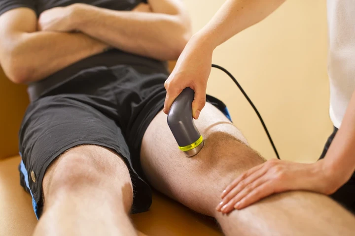
The examination will be carried out by a qualified Sonographer. Warm gel will be applied to the scrotal area, and the Sonographer will move a small transducer across the testicles and surrounding tissues to obtain detailed images.
The scan is non-invasive and should not be painful, although areas of existing tenderness may feel sensitive.
You are welcome to ask questions during your examination.
When do you get the results?
We will send a signed report of our findings to your doctor. You should then arrange an appointment with your GP, who will explain the results.
Your doctor is best placed to discuss the findings because they will have access to your full medical history, examination notes, and any further tests that may be relevant.


Testicular Ultrasound FAQs
At MedicalUltrasound.co.uk, diagnostic accuracy, clinical clarity, and patient reassurance are central to every examination. This FAQ guide covers the most common questions about testicular ultrasound, including when the scan is recommended, what structures are assessed, how Doppler imaging is used, and what happens after your appointment.
The information explains how testicular ultrasound is used to assess the testes, epididymis, and scrotal contents in relation to symptoms such as testicular pain, swelling, lumps, trauma, or fertility concerns. It outlines the role of ultrasound in identifying conditions such as cysts, inflammation, varicoceles, reduced blood flow, or other structural abnormalities that may require urgent assessment or further investigation.
Each question is presented in two parts.
A Short answer for quick reference
An In-depth answer to provide a clinical explanation and context
Whether you have been referred urgently by your GP or are arranging a private scan, this guide is designed to help you understand the purpose of a testicular ultrasound, what to expect during the examination, and how the results support diagnosis, reassurance, or onward care, delivered by experienced diagnostic Sonographers.
What is a testicular ultrasound?
A testicular ultrasound is a scan used to assess the testes and scrotal contents.
In-Depth Answer
Testicular ultrasound uses high-frequency sound waves to produce real-time images of the testes, epididymis, and surrounding scrotal structures. It is commonly used to investigate pain, swelling, lumps, trauma, or fertility-related concerns. The scan is non-invasive, does not use radiation, and provides a detailed assessment of testicular structure and blood flow.
What does a testicular ultrasound assess?
It assesses the testes, epididymis, and scrotal structures.
In-Depth Answer
The scan evaluates testicular size, shape, and internal structure, as well as the epididymis and surrounding tissues. Doppler imaging may be used to assess blood flow. These findings help identify causes of pain, swelling, or lumps and guide further investigation or urgent referral when required.
Can a testicular ultrasound detect testicular cancer?
It can identify suspicious masses, but diagnosis requires further tests.
In-Depth Answer
Testicular ultrasound is very effective at identifying solid masses within the testes, which may raise concern for cancer. However, ultrasound alone cannot confirm a cancer diagnosis. If a suspicious mass is found, urgent referral to urology, blood tests, and sometimes surgery are required. Early imaging is important because testicular cancer is highly treatable when detected early.
Can a testicular ultrasound explain testicular pain?
Yes. It can identify several causes of pain.
In-Depth Answer
Testicular pain may be caused by inflammation, infection, reduced blood flow, cysts, trauma, or other structural abnormalities. Ultrasound helps identify these causes and assess blood supply using Doppler imaging. Some causes of pain may not show clear ultrasound changes, so findings are interpreted alongside clinical assessment.
Can a testicular ultrasound detect infection or inflammation?
Yes. It can identify features of infection or inflammation.
In-Depth Answer
Ultrasound can show enlargement, increased blood flow, and structural changes associated with conditions such as epididymitis or orchitis. These findings support a diagnosis when combined with symptoms, examination, and urine or blood tests. Ultrasound also helps rule out more serious causes of pain, such as torsion.
Can a testicular ultrasound detect torsion?
Yes. It is essential in suspected torsion.
In-Depth Answer
Testicular torsion is a medical emergency caused by the twisting of the spermatic cord, reducing blood flow to the testis. Doppler ultrasound can assess blood flow and support urgent diagnosis. If torsion is suspected clinically, urgent surgical assessment is required, even if imaging is pending. Ultrasound plays a key role in assessment but does not delay emergency care.
Is a testicular ultrasound painful?
No. The scan is usually painless.
In-Depth Answer
Testicular ultrasound is non-invasive and generally well-tolerated. You may feel mild pressure from the probe, especially if the area is tender, but the scan should not be painful. The Sonographer performs the examination sensitively and maintains privacy throughout.
How long does a testicular ultrasound take?
Most scans take around 15 to 20 minutes.
In-Depth Answer
The duration depends on whether both testes are assessed and whether Doppler imaging is required. The scan is performed in real time, and you can return to normal activities immediately afterwards.
Do I need to prepare for a testicular ultrasound?
No special preparation is required.
In-Depth Answer
You can eat and drink normally before the scan and continue taking your usual medications. Wearing comfortable clothing may help. You may be asked to lie down and support the scrotum during the examination.
Will the Sonographer tell me the results during the scan?
Results are provided in a formal written report.
In-Depth Answer
The Sonographer may explain what they are assessing but does not usually provide a diagnosis. A signed clinical report is sent to your GP or referring clinician, who will discuss the findings and advise on next steps.
Can testicular ultrasound detect cysts?
Yes. Cysts are commonly identified on ultrasound.
In-Depth Answer
Ultrasound can identify epididymal cysts, spermatocele, and other benign fluid filled structures. These are often harmless and may require no treatment. The scan helps distinguish cysts from solid masses and guides reassurance or follow up if needed.
Can a testicular ultrasound detect varicoceles?
Yes. Doppler ultrasound is very effective for varicoceles.
In-Depth Answer
Varicoceles are enlarged veins within the scrotum and are a common cause of scrotal discomfort or fertility issues. Ultrasound can assess vein size and blood flow, often using dynamic techniques such as standing or straining. Findings help guide further management or referral.
Can a testicular ultrasound assess fertility problems?
It can provide useful information, but it is not a full fertility assessment.
In-Depth Answer
Ultrasound can assess testicular size, structure, and presence of varicoceles, which may be relevant to fertility. However, fertility assessment also involves semen analysis and hormonal blood tests. Ultrasound findings are interpreted as part of a broader evaluation.
Can ultrasound detect testicular trauma?
Yes. It can assess injury related changes.
In-Depth Answer
Testicular ultrasound can identify bleeding, swelling, rupture, or other structural damage following injury. Early imaging helps guide urgent management and referral when trauma is suspected.
Is a testicular ultrasound safe for repeated scans?
Yes. It does not use radiation.
In-Depth Answer
Ultrasound uses sound waves rather than radiation, making it safe for repeat assessment. This is useful for monitoring known findings or follow-up after treatment or injury.
Can testicular ultrasound detect hydrocele?
Yes. Hydroceles are easily identified.
In-Depth Answer
A hydrocele is a collection of fluid around the testis. Ultrasound can confirm its presence, assess size, and rule out underlying testicular abnormalities. Many hydroceles are benign and require no treatment unless symptomatic.
Can a testicular ultrasound detect hernias?
Sometimes. It may identify inguinal hernias extending into the scrotum.
In-Depth Answer
Ultrasound can sometimes identify hernias that extend into the scrotal sac, particularly during dynamic assessment. A dedicated groin ultrasound may be recommended if hernia assessment is the primary concern.
Can children or adolescents have a testicular ultrasound?
Yes. It is commonly used and safe.
In-Depth Answer
Testicular ultrasound is safe, painless, and commonly used in children and adolescents to assess pain, swelling, or developmental concerns. It avoids radiation exposure and is well tolerated in younger patients.
Do I need a referral for a testicular ultrasound?
A referral is helpful but not always required privately.
In-Depth Answer
Testicular ultrasound is often requested by a GP or specialist, especially when urgent assessment is needed. Private scans may be booked directly, but results should always be shared with your GP to ensure appropriate follow-up.
What happens after my testicular ultrasound?
Your GP will review the report and advise next steps.
In-Depth Answer
After the scan, a signed report is sent to your GP or referring clinician. They will explain the findings and arrange reassurance, monitoring, urgent referral, or further investigation depending on the results.
To book, call: 0141 221 2496 or email[email protected]
To book, call: 0141 221 2496 or email[email protected]
Head Office
MedicalUltrasound.co.uk
Ingram House
227 Ingram Street
Glasgow G1 1DA
Contact
T: 0141 221 2496
Booking line manned
9am to 5pm 7 days a week