
DVT – Deep Vein Thrombosis Ultrasound


DVTDeep Vein ThrombosisUltrasound
Essential information
A DVT ultrasound scan is a specialised vascular ultrasound used to assess the deep veins in the legs for signs of thrombosis (blood clots) in the deep veins (deep vein thrombosis), which can cause swelling, pain, or other symptoms.
Deep vein thrombosis occurs when a blood clot forms within the deep venous system, most commonly in the calf or thigh. These clots can cause pain, swelling, and tenderness in the affected leg.
The scan uses Doppler ultrasound, which allows the Sonographer to assess blood flow, vein compressibility, and the presence of any obstruction within the deep veins. This helps identify whether a clot is present and whether the vein is behaving normally.
A DVT ultrasound is an important investigation because untreated clots can progress or cause complications. Ultrasound may be one of several investigations required, and you should consult your doctor to arrange any further testing or treatment if needed.
Why should I choose MedicalUltrasound.co.uk for my DVT ultrasound scan?
How long is the appointment?
The appointment usually takes around 20- 30 minutes to complete.
How much does it cost?
The cost of this scan is £145.
You can combine this scan with any other scan for an additional £100
Some scans, such as musculoskeletal or vascular imaging, may not be combinable due to specialist requirements.
When should you get a DVT Ultrasound?
You may be advised to have a DVT ultrasound if you have symptoms or examination findings that raise concern for a possible blood clot in the deep veins. This scan helps assess blood flow and the structure of the veins to determine whether a clot is present.
A DVT ultrasound may be appropriate if you have:
- Pain or swelling in one leg, especially in the calf or thigh
- Warmth, redness, or tenderness in the affected area
- Sudden or unexplained swelling of the foot, ankle, or leg
- A feeling of tightness in the calf
- A history of recent long travel or reduced mobility
- Recent surgery, injury, or hospital admission
- A known clotting disorder or family history of DVT
- Use of hormonal medication that may increase the risk of clotting
- A request from your GP or specialist for further investigation
- Follow up after previous DVT or known venous problems
A DVT ultrasound is often the first investigation because it is safe, quick, and provides real-time information using Doppler imaging. It helps your clinician understand whether a clot is present and guides appropriate treatment.
How should you prepare for the scan?
No preparation is required for a DVT ultrasound. You may be asked to remove trousers, tights, socks, or footwear so the Sonographer can easily assess the leg being examined. Wear clothing that can be rolled up or removed comfortably.
What will you experience during the examination?
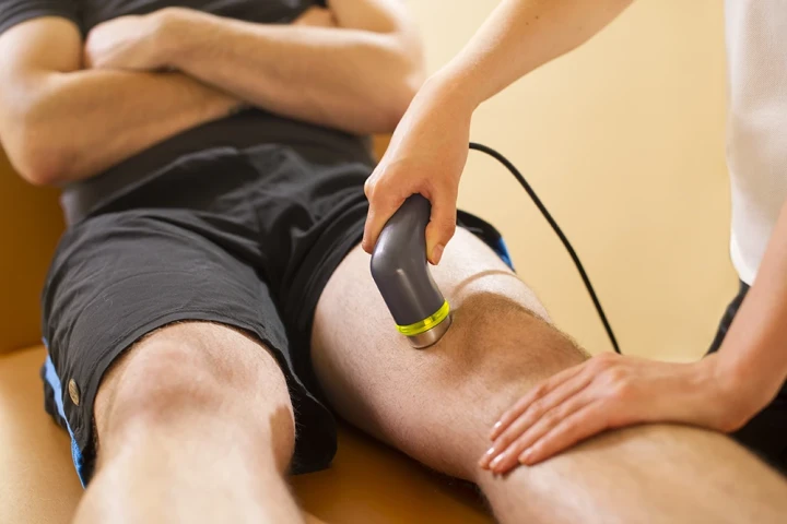
The examination will be carried out by a qualified Sonographer. The procedure is non-invasive and painless. The Sonographer will apply warm gel to your leg and move a small transducer over the deep veins of the calf and thigh.
During the scan, gentle pressure may be applied to assess whether the veins compress normally, which is an important part of detecting a clot.
You may also hear sound from the Doppler function, which allows the Sonographer to assess blood flow through the veins.
You are welcome to ask questions during your examination.
When do you get the results?
We will send a signed report of our findings to your doctor. You should then arrange an appointment with your GP, who will explain the results to you.
Your doctor is best placed to discuss your results because they will have access to your full medical history, blood tests, and any other investigations that may be relevant.


Deep Vein Thrombosis FAQs
What is a DVT ultrasound scan?
It is a scan used to detect blood clots in deep veins.
A DVT ultrasound is a non-invasive vascular scan that assesses the deep veins, usually in the legs, for blood clots. It uses ultrasound and Doppler imaging to evaluate vein compressibility and blood flow. The scan is the first line test for suspected deep vein thrombosis.
What symptoms suggest I might need a DVT ultrasound?
Common symptoms include swelling, pain, and redness in the leg.
DVT ultrasound is often requested if you have unexplained leg swelling, pain, warmth, redness, or tenderness, particularly in one leg. Symptoms may worsen when standing or walking. Because symptoms can be subtle, an ultrasound helps confirm or exclude a clot safely.
How accurate is a DVT ultrasound?
It is highly accurate when performed by trained practitioners.
DVT ultrasound is considered very reliable for detecting clots in the femoral and popliteal veins. Accuracy may be slightly reduced for calf veins, but overall it is the preferred diagnostic test. Repeat scans may be used if symptoms persist or evolve.
Can a DVT ultrasound miss a clot?
It is reliable, but no test is perfect.
Small clots in calf veins or very early clots may be harder to detect initially. If symptoms persist despite a normal scan, your GP may arrange repeat imaging or additional tests. Following preparation and scanning protocols improves diagnostic accuracy.
How long does a DVT ultrasound take?
Most scans take around 20 to 30 minutes.
The duration depends on the extent of the examination and image clarity. The scan is usually completed within half an hour and does not require recovery time. You can resume normal activities immediately afterwards unless advised otherwise.
Do I need to prepare for a DVT ultrasound?
No special preparation is required.
You do not need to fast or stop medications before a DVT ultrasound. It is helpful to wear loose clothing that allows access to the leg being scanned. You will be asked to lie on an examination couch during the procedure.
Can a DVT ultrasound detect pulmonary embolism?
No. It assesses leg veins, not the lungs.
A DVT ultrasound cannot diagnose pulmonary embolism. It assesses the deep veins for clots that may travel to the lungs. If pulmonary embolism is suspected, your GP may arrange other imaging, such as CT pulmonary angiography.
Can a DVT ultrasound be repeated safely?
Yes. It is safe for repeat assessment.
Because ultrasound does not use radiation, it can be repeated if needed. Repeat scans are sometimes used to monitor clot resolution or reassess symptoms. This makes it suitable for ongoing evaluation in selected cases.
Will I get my DVT ultrasound results immediately?
The formal results are provided in a written report.
The Sonographer may explain what they are assessing during the scan, but the official findings are documented in a signed report. This is sent to your GP or referring clinician, who will discuss the results and next steps with you.
What happens if a blood clot is found on an ultrasound?
Your GP will arrange urgent treatment or referral.
If a clot is detected, the report will document its location and extent. Your GP may start anticoagulant treatment, arrange urgent referral, or organise further tests depending on the findings and your clinical situation. Prompt management reduces the risk of complications.
Can a DVT ultrasound detect clots above the knee?
Yes. A standard DVT ultrasound assesses veins in the calf and thigh.
A DVT ultrasound typically examines the deep veins of the lower leg and thigh, including the femoral and popliteal veins. This allows detection of clots above and below the knee. In some cases, a more extensive vascular study may be requested to assess additional veins. Your GP will decide the appropriate scan based on symptoms and clinical risk.
Will a DVT ultrasound show if the clot is new or old?
It can suggest this, but timing is interpreted clinically.
Ultrasound can identify features that suggest whether a clot is acute or chronic, such as vein compressibility, clot appearance, and vein wall changes. However, ultrasound cannot always determine the exact timing. Your GP or specialist will interpret the findings alongside your symptoms, medical history, and any previous imaging.
Can I have a DVT ultrasound if my leg swelling comes and goes?
Yes. Intermittent symptoms can still be assessed.
Fluctuating swelling does not rule out a blood clot. A DVT ultrasound can assess the deep veins even if symptoms are not constant. The scan may also identify other causes of swelling, such as fluid collections or soft tissue changes, helping guide further management.
Is a DVT ultrasound useful after long flights or travel?
Yes. Travel-related immobility increases clot risk.
Long-haul flights, prolonged car journeys, or periods of immobility can increase the risk of deep vein thrombosis. If symptoms such as leg pain, swelling, or redness develop after travel, a DVT ultrasound is commonly used to assess the deep veins and exclude or confirm a clot.
Can a DVT ultrasound help if I have varicose veins?
It assesses deep veins, not superficial varicose veins.
A DVT ultrasound focuses on the deep venous system, which is responsible for most clinically significant clots. Varicose veins affect superficial veins and require a different type of vascular ultrasound if assessment is needed. Your GP may request a separate scan if superficial venous disease is suspected.
Does a DVT ultrasound check both legs?
Usually, the affected leg, unless both are symptomatic.
In most cases, the scan is performed on the leg with symptoms. If both legs are affected or if your GP requests bilateral imaging, both sides can be assessed. The decision is based on symptoms, risk factors, and clinical judgment.
Can medication affect the results of a DVT scan?
Blood thinners may alter appearance, but do not prevent assessment.
Anticoagulant medication can change how a clot appears over time, but ultrasound can still assess vein compressibility and blood flow. The scan remains useful for evaluating treatment response or ongoing symptoms. Your GP will interpret findings in the context of your medication history.
Is it normal to feel pressure during the scan?
Yes. Compression is an essential part of the test.
During a DVT ultrasound, gentle pressure is applied with the probe to check whether the veins compress normally. Non-compressible veins may indicate a clot. This pressure is usually well tolerated and does not cause harm, though mild discomfort can occur over tender areas.
Can a DVT ultrasound show other causes of leg pain or swelling?
Yes. Alternative findings may be identified.
While the primary aim is to detect blood clots, a DVT ultrasound may also reveal other causes of symptoms. These include fluid collections, Baker’s cysts, muscle injury, or soft tissue swelling. Identifying alternative explanations can help guide appropriate care.
Is a DVT ultrasound safe during pregnancy?
Yes. It is safe and commonly used in pregnancy.
Ultrasound does not use radiation and is safe during pregnancy. Because pregnancy increases the risk of blood clots, DVT ultrasound is frequently used to assess leg symptoms promptly and safely. It allows accurate assessment without risk to the baby.
To book, call: 0141 221 2496 or email[email protected]
To book, call: 0141 221 2496 or email[email protected]
Head Office
MedicalUltrasound.co.uk
Ingram House
227 Ingram Street
Glasgow G1 1DA
Contact
T: 0141 221 2496
Booking line manned
9am to 5pm 7 days a week